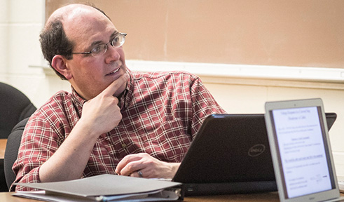
Dr. David Tees
By Amanda Biederman
NQPI editorial intern
Ohio University Professor of Physics & Astronomy and Nanoscale & Quantum Phenomena Institute member David Tees is investigating the mechanical properties of cancer stem cells to elucidate how they propagate throughout the body.
It is well known that tumor cells can enter the blood and form secondary tumors in other tissues. However, the mechanism behind this process is not fully understood. Tees is working with Monica Burdick, an OHIO professor of Chemical and Biomolecular Engineering, to characterize how breast cancer cells travel through vessels. Tees’ team includes current and former graduate students Young Eun Choi, Amina Alipour, Aaron Burdette and Pooja Chopra.
During development, stem cells differentiate (mature) into discrete cell types that are normally fixed for the organism’s lifetime. However, when cancer cells become mesenchymal, they can dedifferentiate, taking on stem-like properties and often redifferentiating as a secondary tumor.
“Once the cell goes through this transition, its mechanical properties might change,” Tees said. “These cells are more aggressive … and potentially more able to propagate in the body at another site.”
Tees hypothesized that mesenchymal cells mimic healthy leukocytes, allowing them to circulate undetected. Metastatic cells are initially large and rigid, but by altering their mechanical properties, they may be able to pass through pulmonary capillaries. Tees’ team uses two methods to measure cancer stem cell flow behavior.
In the first method, Tees’ team observes the movement of cancer cells into a micropipette under pressure. They have characterized the relationship between stress (pressure) and strain (cell deformation).
The group is developing a second method based on kinematics that utilizes microfluidics to track cell movement. In this system, a cell sample travels through a series of tapered vessels to a channel that is narrower than the cell diameter. They measure the time cells take to deform and enter the narrow section. Although his group monitors flow microscopically, Tees hopes to develop a system with electronic sensors, which would allow clinicians to collect information on a patient’s risk of metastasis.
“Watching these cells go by is … a complex skill and labor-intensive,” Tees said. “An electronic measurement that tracks the movement of cells … would be a more practical device … that’s usable for a non-expert.”


Comments