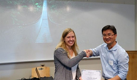
HTC Neuroscience student Samantha Selhorst with Neuroscience Program Director Dr. Daewoo Lee at Neuroscience Research Day (photo: Muhammad Fauzi)
By Amanda Biederman
Rare diseases are often understudied, but Honors Tutorial College Neuroscience student Samantha Selhorst is working to characterize the pathology of arteriovenous malformation (AVM) under the direction of Biological Sciences professor Dr. Corinne Nielsen. Selhorst was recently awarded first prize for her thesis work in the undergraduate competition at Neuroscience Research Day.
AVM is a rare condition that affects less than 1 percent of the U.S. population. For this reason, the pathology of AVM is not well understood, and treatment options are often limited. AVM is characterized by the development of blood vessels that disrupt the normal capillary network in the brain. AVM tangles often promote brain hemorrhages that cause internal bleeding and can lead to death in patients.
“A lot of patients develop enlarged tangled blood vessels that sit on or within the brain. So if it’s on the cortex, a neurosurgeon can fairly easily go in a do a surgical resection,” Selhorst said. “But if it’s deep within the cortex, surgeons can’t go in to get it because they would cause damage. The patients with more severe AVMs often don’t have any treatment options.”
To understand the pathology of AVM, Selhorst is using a mouse model to examine the prevalence of pericytes in different regions of the brain during AVM progression. Pericytes are a type of cell that enwrap endothelial cells lining blood vessels. These cells are important for maintenance of the blood-brain barrier, which helps separate the brain from the rest of the body’s circulatory system. However, the exact functional role of pericyte cells is not well understood.
Selhorst used immunostaining to measure the prevalence of pericytes in the cerebellum, brainstem and cortex. She found that in mice exhibiting AVM, pericyte abundance was elevated in both the cerebellum and the cortex.
In a future study, Selhorst hopes to better understand how pericytes become more abundant during disease progression. She plans to look at the number, shape and size of these cells during disease, and she said she is also interested in the signaling pathway that may stimulate pericyte abnormalities
Selhorst said her project stemmed from discussions with Nielsen, which she carried out as part of the HTC program. Ultimately, this work may contribute to the medical community’s understanding of how this cell type may be involved in the progression of this rare medical condition.
“(Dr. Nielsen and I) were talking about AVM in general and the neurovascular unit in that system, and somehow pericytes were brought up,” Selhorst said. “I really thought it was interesting that (pericytes) are considered part of the vascular system, but they play such an important role for the nervous system.”


Comments