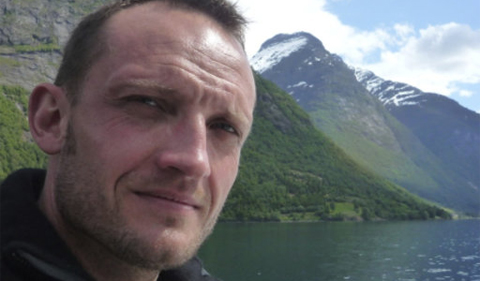
Ohio University’s Chemistry & Biochemistry Colloquium Series presents Dr. Cyril Ruckebusch on “A chemometric view of super-resolved fluorescence microscopy imaging” on Monday, Oct. 29, from 4:10 to 5 p.m. in Clippinger 194.
Ruckebusch is with Université de Lille in France.
Abstract: Super-resolution wide-field fluorescence microscopy can provide structural information at the nanoscale and dynamic insight about biological processes in live cell samples. In general, the available information in super-resolution images is related to the density of emitters, with more emitters leading to more information. One of the strategies for obtaining a high spatial resolution is based on the sequential imaging and localization of sparse subsets of blinking fluorophores distributed over thousands of images, resulting in a high-density image of their positions and intensities. However, to obtain a high spatial resolution on short time sampling, and potentially probe dynamic processes in live cells, this principle must be extended to the analysis of high-density of emitters distributed over a few tens of movies frames only. As many emitters are simultaneously active, their emissions strongly overlap and single-emitter fitting methods collapse. Thus, analyzing high-density super-resolution data, the development of new methods remains a challenging issue for dynamic imaging and faster super-resolution experiments.
The core of our approach is the SPIDER algorithm for SParse Image DEconvolution and Reconstruction. This approach tackles the image deconvolution problem in a penalized regression framework with a combination of a sparseness and an inter-frame penalty. Sparseness of the fluorophore spatial distribution is obtained with an L0-norm penalty on estimated fluorophore intensities, effectively constraining the number of fluorophores per frame. Simultaneously, continuity of the fluorophore localizations is obtained penalizing the total numbers of pixel status changes between successive frames. We show that more accurate estimates of the final super-resolution images of cellular structures can be obtained.
Additionally, a high-density of the fluorophore labelling translates into the presence of significant auto-fluorescence background and strong bleaching of the fluorophores, masking the blinking. Preprocessing is thus required at a very first step towards a super-resolution image, and combination of spatial and temporal approaches might be required. We show that with proper pre-processing of the data, super-resolution images have better contrast and better resolution.
The host is Dr. Peter Harrington, Professor of Chemistry & Biochemistry at Ohio University.


Comments