

Dr. Jixin Chen
Dr. Jixin Chen, Assistant Professor of Chemistry & Biochemistry and Nanoscale & Quantum Phenomena Institute member, published multiple journal articles in 2017.
Chen has developed a statistical computer program to help researchers account for the blurring effect in single-molecule Förster resonance energy transfer (smFRET) measurements. Single-molecule FRET is a classic microscopy method that uses fluorescent dyes to measure the distance between two points within a molecule. The technology can be used to track temporal changes in molecular structure, such as those of a membrane transport protein that alters its conformation in open or closed states to allow molecules to pass through it.
Joseph R. Pyle, Jixin Chen,* Photobleaching of YOYO-1 in Super-Resolution Single-DNA Fluorescence Imaging. BJNano. 2017, 8, 2296-2306. doi:10.3762/bjnano.8.229.
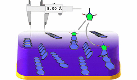
Abstract: Super-resolution imaging of single DNA molecules via point accumulation for imaging in nanoscale topography (PAINT) has great potential to visualize fine DNA structures with nanometer resolution. In a typical PAINT video acquisition, dye molecules (YOYO-1) in solution sparsely bind to the target surfaces (DNA) whose locations can be mathematically determined by fitting their fluorescent point spread function. Many YOYO-1 molecules intercalate into DNA and remain there during imaging, and most of them have to be temporarily or permanently fluorescently bleached, often stochastically, to allow for the visualization of a few fluorescent events per DNA per frame of the video. Thus, controlling the fluorescence on–off rate is important in PAINT. In this paper, we study the photobleaching of YOYO-1 and its correlation with the quality of the PAINT images. At a low excitation laser power density, the photobleaching of YOYO-1 is too slow and a minimum required power density was identified, which can be theoretically predicted with the proposed method in this report.
Gregory Deye, Juvinch R. Vicente, Shawn M. Dalke, Selma Piranej, Jixin Chen, Jacob W. Ciszek, The Role of Thermal Activation and Molecular Structure on the Reaction of Molecular Surfaces. Langmuir 2017, 33(33), 8140-8146
Abstract: Though surface modifications of organic thin films dramatically improve optoelectronic device performance, chemistry at organic surfaces presents new challenges that are not seen in conventional inorganic surfaces. This work demonstrates that the subsurface of pentacene remains highly accessible, even to large adsorbates, and that three distinct reaction regimes (surface, subsurface, and bulk) are accessed within the narrow thermal range of 30–75 °C. Progression of this transition is quantitatively measured via polarization modulation infrared reflection absorption spectroscopy, and atomic force microscopy is used to measure the thin-film morphology. Together, they reveal the close relationship between the extent of the reaction and the morphology changes. Finally, the reaction kinetics of the pentacene thin film is measured with a series of adsorbates that have different reactivity and diffusivity in the thin film. The results suggest that reaction kinetics in the thin film is controlled by both the reactivity and the adsorbate diffusivity in the thin-film lattice, which is very different than the traditional solution kinetics that is dominated by the chemical activation barriers. Combined, these experiments guide efforts toward rationally functionalizing the surfaces of organic semiconductors to enable the next generation of flexible devices.
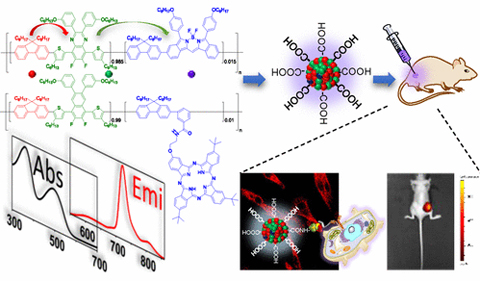
Chi-Shiang Ke, Chia-Chia Fang, Jia-Ying Yan, Po-Jung Tseng, Joseph R. Pyle, Chuan-Pin Chen, Shu-Yi Lin, Jixin Chen, Xuanjun Zhang, Yang-Hsiang Chan, Molecular Engineering and Design of Semiconducting Polymer Dots with Narrow-Band, Near-Infrared Emission for in Vivo Biological Imaging. ACS Nano 2017, 11(3), 3166-3177
Abstract: This article describes the design and synthesis of donor–bridge–acceptor-based semiconducting polymer dots (Pdots) that exhibit narrow-band emissions, ultrahigh brightness, and large Stokes shifts in the near-infrared (NIR) region. We systematically investigated the effect of π-bridges on the fluorescence quantum yields of the donor–bridge–acceptor-based Pdots. The Pdots could be excited by a 488 or 532 nm laser and have a high fluorescence quantum yield of 33% with a Stokes shift of more than 200 nm. The emission full width at half-maximum of the Pdots can be as narrow as 29 nm, about 2.5 times narrower than that of inorganic quantum dots at the same emission wavelength region. The average per-particle brightness of the Pdots is at least 3 times larger than that of the commercially available quantum dots. The excellent biocompatibility of these Pdots was demonstrated in vivo, and their specific cellular labeling capability was also approved by different cell lines. By taking advantage of the durable brightness and remarkable stability of these NIR fluorescent Pdots, we performed in vivo microangiography imaging on living zebrafish embryos and long-term tumor monitoring on mice. We anticipate these donor–bridge–acceptor-based NIR-fluorescent Pdots with narrow-band emissions to find broad use in a variety of multiplexed biological applications.
Joseph R. Pyle, Kurt W.E. Sy Piecco, Juvinch R. Vicente, Jixin Chen,* Optical Genome Mapping. Encyclopedia of Chemical Processing. accepted.


Comments