
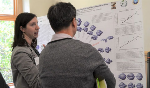
Ph.D. student Catherine Early presents her research to Biological Sciences professor Daewoo Lee.
The brain recounts a unique story. The size, shape, and each fold along the surface reveal a narrative about how an animal lives, as well as how its ancestors have evolved over millions of years.
But what about animals that have died, with only a skeleton or fossilized skeleton remaining? Although the brain may be long gone, researchers often gather information by examining the cavity within the skull.
Catherine Early, a fifth-year Ph.D. student in Biological Sciences, is assessing this indirect method used for inference of avian brain morphology. Early was recently awarded first prize (tied with Ph.D. student Mary Gemmel) for the best graduate student poster at Neuroscience Research Day.
Researchers draw conclusions about brain morphology by creating digital endocasts, computer-generated models of the skull cavity, using computerized tomography (CT) scanning. Next, they analyze the three-dimensional rendering piece by piece, characterizing the shapes of different brain regions as they appear on the external surface of the endocast. But according to Early, it’s difficult to know if these endocasts tell the full story.
“CT scanning has become really prevalent in our field,” Early said. “When you make an endocast that reflects the brain, the assumption is that because it looks like the brain, the size of an endocast structure correlates with the size of the brain structure.… The point of this project is to test that assumption.”
Early, who works under the supervision of Biomedical Sciences professor Dr. Larry Witmer, is creating digital endocasts from species with known brain morphologies and recorded behavioral traits. Using the endocasts, she is calculating the relative sizes of two endocast regions – the optic lobe and the Wulst – that overlie brain structures involved in visual pathways. Next, she compares her data with previously published data on the brain structures.
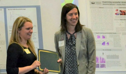
Catherine Early (right) and Mary Gemmel (left) both won first prize for graduate student posters at Neuroscience Research Day.
Early said she plans to incorporate approximately 50 species of birds into her dataset and that she is working to incorporate a broad taxonomic array. Thus far, Early said her analyses indicate that the connection between brain and endocast measurements holds true.
Ultimately, Early said this study will help researchers who want to learn about the behaviors and evolutionary histories of extinct birds who have only left fossilized remains behind.
“(If) you can use the surface area in extinct birds to predict the volume, (you can) incorporate the volume into the analyses that have already been done in extant birds,” Early said. “Incorporating them could help me figure out … when (certain traits) evolved, how much variation there was in the fossil record, (and) the normal baseline throughout evolution. That’s the ultimate goal.”
- More information about the Neuroscience Program at Ohio University.


Comments